
The Sentence That Starts So Many Appointments
“He’s had this little cough for a couple of weeks. I think it’s just a leftover from kennel cough.”
Or: “She coughs when she gets excited. Probably nothing.”
Most coughs in dogs and cats are, in fact, minor and self-limiting. A subset are not. The difficulty is that the early history often sounds identical. The difference usually appears in the details of the exam, the timeline, and the quiet findings that do not make it into the Facebook post about “home remedies for cough.”
Here are three cases that still sit with me. Each began with the same understated description. Each ended in a different place. Together they illustrate why I still treat every persistent cough as a chart that has not finished speaking.
Case 1: The Young Labrador With the “Kennel Cough That Wouldn’t Quit”
Max was a two-year-old Labrador who had boarded three weeks earlier. A harsh, honking cough started four days after he came home. The owner waited the usual ten days, then another week, then brought him in because the cough was still present and now occasionally ended in a gag.
On exam Max was bright, afebrile, and eager to greet everyone. Lung sounds were clear. Tracheal sensitivity was mild. The history fit infectious tracheobronchitis, but the duration was longer than expected.
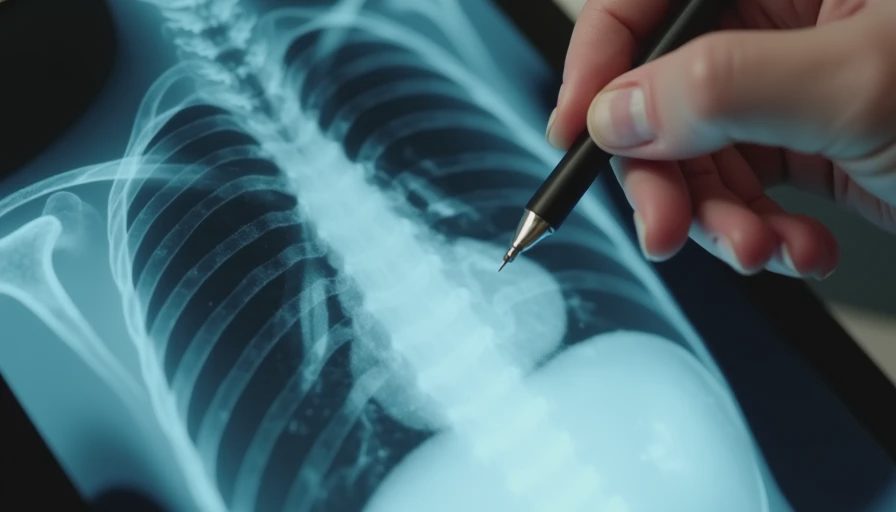
We took radiographs. The films showed a soft-tissue opacity at the carina that had no business being there in a young dog with a simple infectious cough. Bronchoscopy followed. A small plant awn was lodged in the right mainstem bronchus, surrounded by early granulation tissue. Removal was straightforward. The cough resolved within forty-eight hours.
Lesson I still use: Infectious coughs improve or resolve on a predictable timeline. When the timeline stretches and the dog still looks systemically well, look for a foreign body or other mechanical cause before simply extending antibiotics another round.
Case 2: The Older Cat Whose Cough Was “Only After Naps”
Luna was a twelve-year-old domestic shorthair. Her owner described a soft, occasional cough that seemed to happen when Luna stretched after sleeping. No weight loss, normal appetite, no open-mouth breathing at home. The referring note said “possible mild asthma.”
In the exam room Luna’s resting respiratory rate was 36. Subtle increased bronchovesicular sounds were present dorsally. Chest films showed a diffuse bronchial pattern and mild hyperinflation. Bloodwork was largely unremarkable except for a mild eosinophilia.
We treated for presumptive allergic airway disease and the cough improved—but not completely. A month later the resting rate was still elevated and the owner reported two episodes of what looked like brief “panting” at night. We repeated imaging and added a pro-BNP. The heart size was borderline; the pro-BNP was elevated. An echocardiogram confirmed early hypertrophic cardiomyopathy with left atrial enlargement. The cough was cardiac.
Lesson I still use: Cats hide respiratory effort better than dogs. A “soft occasional cough” plus any rise in resting respiratory rate deserves both airway and cardiac differentials until proven otherwise. Asthma and heart disease can look identical at home.
Case 3: The Middle-Aged Beagle Whose Cough Came With a Shrug
Daisy was seven. The cough had been present for two months—dry, intermittent, worse at night. The owner had tried a course of antibiotics from a previous clinic and a cough suppressant. Nothing changed much. Daisy still wanted her walks.
Exam showed good body condition, mild periodontal disease, and a subtle grade 2/6 left-sided systolic murmur that the owner had never been told about. Lung sounds were normal. Radiographs revealed a moderately enlarged left atrium and mild pulmonary venous congestion. No obvious pneumonia or mass.
Echocardiography confirmed myxomatous mitral valve disease with enough volume overload to produce early congestive changes. We started a diuretic and a heart-failure protocol. The nighttime cough disappeared within five days.
Lesson I still use: A new or progressive cough in a middle-aged or older dog is cardiac until the chart says otherwise. The murmur is not always loud. The owner may not know it exists. Nighttime cough is a classic early sign of elevated left-atrial pressure, and it is easy to dismiss as “something she does when she lies down.”
The Common Thread
In each case the initial description was ordinary. The differentiating data came from:
Precise timeline and progression
Resting respiratory rate and effort
Careful auscultation (including the heart)
Imaging timed to the history rather than delayed by “wait and see”
Willingness to revisit the working diagnosis when the expected improvement did not appear
I still start every cough appointment with the same open posture: most of these are minor. Then I let the individual chart tell me whether this one belongs in the minority that needs more.
What Owners Can Do Before the Appointment
Count the resting respiratory rate at home while the pet is deeply asleep.
Note whether the cough is productive or dry, worse at night or with activity, and whether it is changing.
Record a short video.
Bring any previous records so we can see the timeline clearly.
Those details shorten the path from “just a cough” to the correct next step.
The three animals above all went home and did well once the real problem was identified. The ones that stay with me are the ones that almost stayed in the “probably nothing” column a little too long. That is why I still listen to every cough as if the chart has more to say.
Let the chart do the talking.




No letters yet — be the first to write.